
Breaking Down Sponge Shapes
Jul 21st 2026
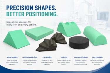
Walk into any well-equipped radiology department and you'll find a collection of foam sponges in an assortment of shapes, sizes, and angles. To the uninitiated, they look like random geometric shapes. To the experienced technologist, each one is a precision tool designed for specific anatomical positioning challenges.
Positioning sponges serve two connected purposes: ensuring the patient is in the correct anatomical position for a diagnostic-quality image and keeping the patient comfortable enough to hold that position without moving during the exposure. Get both right, and you reduce retakes, minimize patient dose, and improve diagnostic accuracy. The key is knowing which sponge to reach for and when.
Wedge Sponges: The Workhorse of Positioning
Wedge sponges are available in 15°, 30°, 45°, and other angles and are the most versatile positioning tools in radiology. Their angled surface provides controlled tilt without requiring the patient to actively hold an awkward position.
45-degree wedges: The most used angle. Ideal for oblique views of the chest, abdomen, and extremities. A 45-degree wedge placed under the patient's hip creates a perfect oblique pelvis position. Under the shoulder, it opens the glenohumeral joint for AP oblique views.
30-degree wedges: Used for gentle oblique positioning where 45 degrees is too steep. Common in lateral decubitus chest positioning support, cervical spine obliques, and gentle rotation of the thorax.
15-degree wedges: Subtle angle adjustments for fine-tuning positions. Used under the knee for slight flexion during AP knee views, or under the ankle for AP mortise views where a small degree of rotation is needed.
Wedge sponges with unique angles are designed to provide artifact-free viewing with no lines on the radiology image, combined with the stability needed to hold the position throughout the exposure.
Rectangular Blocks: Elevation and Support
Rectangular positioning blocks provide flat, stable elevation at consistent heights. They're used when you need to raise an anatomical part to center it in the x-ray beam, support a body part at a specific height, or fill the gap between the patient's anatomy and the table or image receptor.
Thin rectangles (1-2 inches): Placed under the wrist for PA hand views, under the forearm for lateral elbow positioning, or under small body parts to center them within the beam field.
Medium rectangles (3-4 inches): Support under the knee for lateral knee views, under the shoulder for AP external rotation views, or as gap fillers in cross-table lateral positioning.
Large rectangles (5+ inches): Used for significant elevation and positioning a patient's upper body for upright abdominal views when the department lacks a wall bucky, or supporting the torso during decubitus positioning.
Bolsters: Cylindrical Support
Bolster sponges are cylindrical or semi-cylindrical shapes that conform to and support the natural curves of the body. Their rounded profile makes them particularly useful for positioning extremities and providing comfortable support under lordotic curves.
Under the knees: A bolster placed under a supine patient's knees reduces lumbar lordosis and improves patient comfort during AP lumbar spine and AP abdomen examinations. Comfortable patients hold still and that reduces retakes.
Under the neck: A cervical bolster supports the natural cervical lordosis during supine head and neck positioning, maintaining alignment without hyperextension.
Extremity support: Small bolsters cradle the forearm, wrist, or ankle in specific rotational positions, preventing the limb from rolling during the exposure.
Specialty Shapes
Beyond the standard wedges, rectangles, and bolsters, Techno-Aide manufactures specialty sponge shapes designed for specific examinations:
Head sponges: Contoured to cradle the skull during various cranial projections, maintaining head position without manual holding.
Finger and hand sponges: Stepped or angled surfaces designed to fan out the fingers or angle the hand for specific views without requiring the patient to maintain muscular effort.
Material Matters: Closed-Cell Foam
Positioning sponges must have minimal artifacts on the radiographic image. Techno-Aide's positioning sponges use closed-cell foam that is entirely radiolucent. Unlike open-cell foam, which absorbs fluids and harbors bacteria, closed-cell foam is inherently non-porous, resisting moisture absorption and simplifying infection control.
This material distinction has significant infection control implications. In a department where positioning sponges contact multiple patients throughout the day, a non-porous surface that can be fully wiped clean between uses is a meaningful advantage over materials that absorb body fluids and disinfectant solutions.
Building a Complete Sponge Inventory
A well-rounded sponge collection for a general radiography room should include a mix of wedges in at least two angles (45° and 30°), rectangular blocks in two or three heights, at least one bolster for lumbar support, and specialty shapes appropriate to the examinations performed in that room. For specialized rooms like pediatric suites, orthopedic clinics, trauma bays, the sponge selection should be tailored to the exam mix.
Techno-Aide supplies the full range of positioning sponge shapes in multiple sizes, with options for coated and non-coated surfaces finishes. Our team can help you build a sponge inventory matched to your department's exam volume and specialty mix.